
Structure of a Muscle Cell
This follows general information about the structure of muscle.
Skeletal muscles consist of 100,000s of muscle cells (also known as 'muscle fibres' and 'muscle fibers'). They perform the functions of the specific muscle of which they are a part.
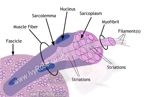
Above: Diagram of a Muscle Cell
|
|
||||||||||||||
|---|---|---|---|---|---|---|---|---|---|---|---|---|---|---|---|
Above: Diagram of a Muscle Cell |
|||||||||||||||
The diagram above shows some detail of the structure of a muscle cell. It illustrates the distinctive structure of muscle cells, including striated myofibrils (components of muscle cells only).
For further information about the parts of the muscle cell labelled above:
It is also important to remember that organelles essential for all cells (not all of which are illustrated in the diagram above) are also present in muscle cells. For more about the contents of cells generally see animal cells.
General components of Muscle Fibres/cells:
The components of skeletal muscle cells that are specific to muscle tissue are myofibrils.
- Each muscle fibre ('muscle cell') is covered by a plasma membrane sarcolemma.
- Tunnel-like extensions from the sarcolemma pass through the muscle fibre from one side of it to the other in transverse sections through the diameter of the fibre. These tunnel-like extensions are known as transverse tubules ('T Tubules') - not shown in diagram above.
- The nuclei of muscle fibres ('muscle cells') are located at the edges of the diameter of the fibre, adjacent to the sarcolemma. As illustrated, a single muscle fibre may have many nuclei.
- Cytoplasm is present in all living cells.
The cytoplasm present is muscle fibres (muscle cells) is called sarcoplasm. - The sarcoplasm present in muscle fibres contains very many mitochondria, which are the energy-producing units within the cell. These mitochondria produce large amounts of a chemical called 'Adenosine Triphosphate', which is usually referred to in abbreviated form as 'ATP'. (The cellular activities for which ATP is required include contracting muscles, moving chromosomes during cell division, moving structures with cells, transporting substances across cell membranes, and synthesizing larger molecules from smaller ones. To understand the function of ATP for the actions of muscle fibres / cells, remember that ATP is necessary for muscle contraction, and is produced by the mitochondria within the muscle
- Sarcoplasmic reticulum is a network of membrane-enclosed tubules similar to smooth endoplasmic reticulum (SER). Sarcoplasmic reticulum is present in muscle fibres/cells and extends throughout the sarcoplasm of the cell. The function of the sarcoplasmic reticulum is to store calcium ions, which are necessary for muscle contraction.
- Myoglobin is also present in the sarcoplasm of muscle fibres/cells. This is a reddish pigment that not only results in the distinctive colour of skeletal muscle, but also stores oxygen until it is required by the mitochondria for the ATP.
More specialised components of Muscle Fibres (Muscle Cells):
The components of skeletal muscle cells that are specific to muscle tissue are called myofibrils. These are cylindrical structures (see above) that extend along the complete length of the muscle fibre/cell.
Each myofibril consists of two types of protein filaments called 'thick filaments', and 'thin filaments'. These two types of filament have different structures, as shown on the page of diagrams of muscle filaments.
In simple terms, the thick filaments and the thin filaments within myofibrils overlap in a structured way, forming units called sarcomeres.
Sarcomeres are sections of myofibril that are separated from each other by areas of dense material called 'Z discs'. Sarcomeres are also described in terms of the bands / zones within which one or both of the two filaments occur.
These bands / zones are illustrated in the diagram below:
- The 'A band' is a relatively darker area within the sarcomere that extends along the total length of the thick filaments.
- The 'H zone' is at the centre of the A band of each sarcomere. As shown below, this is the region in which there are only thick filaments, and no thin filaments.
- The 'I band' is the region between adjacent A bands, in which there are only thin filaments, and no thick filaments. (Each I band extends across two adjacent sarcomeres.)
In the diagram below the Z discs are represented by the zig-zag lines that form the boundaries between adjacent sarcomeres.
Above: Diagram of one sarcomere
(within a muscle cell)

|
Diagram of one sarcomere (within a muscle cell). |
The next page describes the structure of muscle filaments ...


